Onlay bone grafting at Clínica Loyola in Bogotá, Colombia, rebuilds jawbone volume when there is too little bone to support a dental implant. Planning uses 3D tomography and patient-specific, 3D-printed surgical guides. Oral and maxillofacial surgeon Dr. Juan Carlos Farré has over 30 years of experience and is the clinic’s scientific director.
The procedure adds a bone block to the upper or lower jaw. It may be considered after tooth loss, gum disease, infection, or trauma causes bone loss. Long-term use of removable dentures can also lead to gradual loss of jawbone.
Clínica Loyola holds JCI accreditation and ISO 9001 certification. The center has its own operating rooms. Dr. Farré specializes in oral and maxillofacial surgery, dental implants, and bone regeneration.
3D planning and graft materials
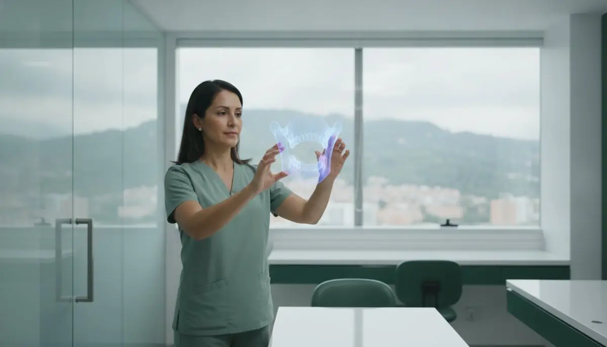
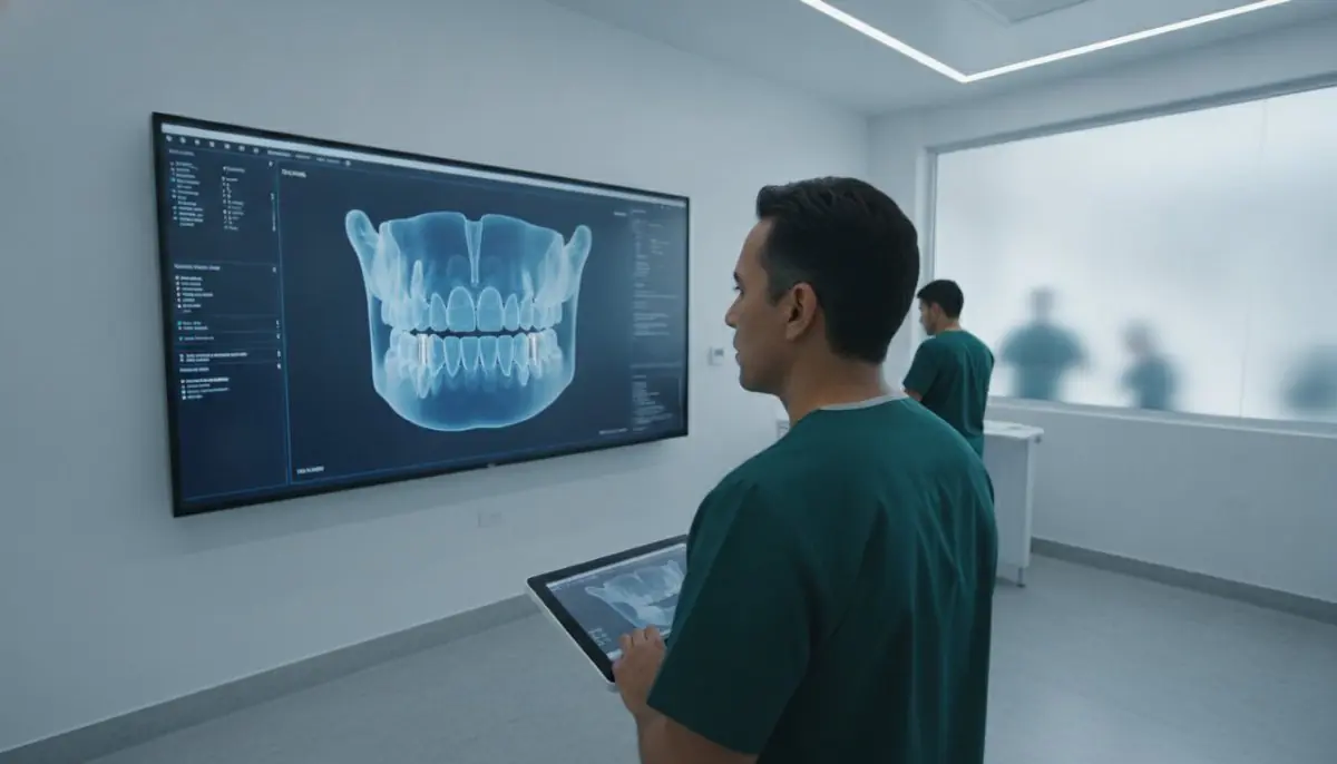
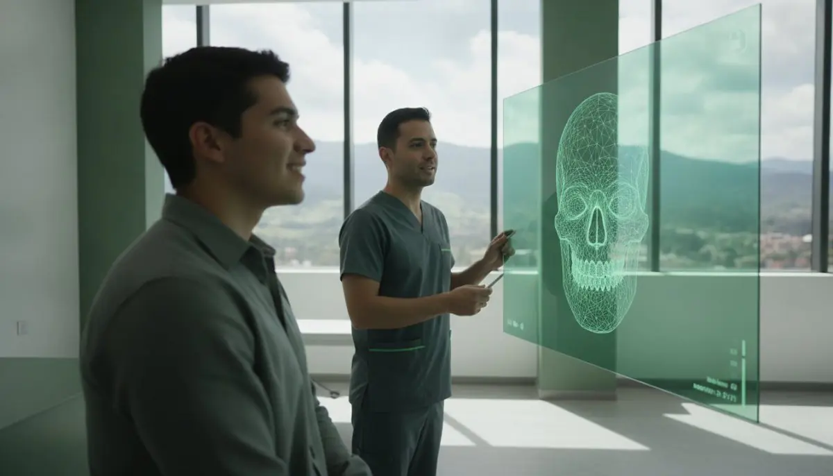
The team uses 3D tomography to assess the area and plan graft placement. Graft material is selected based on the defect, the bone volume needed, tissue condition, and the planned dental restoration. Options include the patient’s own bone, processed tissue-bank graft, or synthetic biomaterials designed to support bone formation.
Before surgery, patients may need blood tests to check glucose and blood clotting. The team reviews medical history, checks medications, and gives fasting instructions. These steps help plan care around each patient’s health needs.
Bone graft healing and follow-up
The graft acts as a temporary scaffold while the body forms new bone around the material. Healing follows an individual biological timeline. Regular visits let the team monitor graft integration and plan the next steps in oral rehabilitation.
Follow-up may also involve orthodontics and post-operative checks, depending on the treatment plan. Patients can request a consultation through Bookimed to discuss whether onlay bone grafting may fit their needs.


 The clinic is not accepting new requests
 Average answering time – 5 minutes
